 Our most recent program focuses on targeted drug delivery of chemotherapy agents and the cell apoptosis (programmed cell death) that occurs as a result of successfully killing the cancer cell.
A major aspect of this work involves the study of poly(amidoamine) (PAMAM) dendrimers as drug transport agents. The polymeric particle used as the platform for drug transport is the PAMAM dendrimer. A space-filling model of a generation 4 polymer molecule is shown here.
The additional functionality needed for selective drug delivery is covalently attached to the particle. This includes targeting and imaging groups as well as the drug to be delivered.
We are working to understand a number of aspects of this system:
- the structure of the functionalized drug transport agents
- the mechanism of the dendrimer incorporation into the cell
- the mechanism of drug release
- the ultrastructural aspects of cell apoptosis
Initial aspects of our work were published in Langmuir.
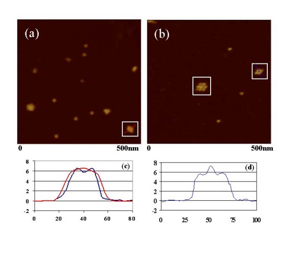
A comparison of PAMAM dendrimers imaged with a commercial AFM tip and a single-wall carbon nanotube AFM tip grown in our lab is shown below. In panel a, the clusters of three,four, and five dendrimers are apparent, however, there is not direct resolution of the individual particles. In panel b, the individual dendrimers are directly visualized. A comparison of the two 4 dendrimer clusters is shown in panel c. Panel d give a line scan of the seven dendrimer cluster highlighted in panel b.
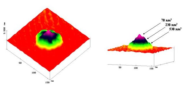
We have also studied covalently linked clusters of dendrimers known as tectodendrimers. Tectodendrimers consists of a PAMAM dendrimer core surrounded by a lower generation dendrimer that are covalently linked to the central dendrimer. In the image shown below, a G7 dendrimer linked to ~11 G5 dendrimers is adsorbed onto a mica surface. AFM analysis allows the number of G5 dendrimers linked to the G7 core to be assessed. One advantage of the tectodendrimer architecuture is the ability to make each individual dendrimer perform a separate function.
This image is in many ways a wonderful technical accomplishment. We used single-wall carbon nanotube probes to directly measure the core to shell dendrimer ratio in this unique nanodevices. However, my good colleague Prof. Neil Marsh saw things somewhat differently. His rather unique view, posted on my door in the mid of night, is reprinted here for your amusement.
We are very actively pursuing the effects the nanodevices have on cell apoptosis. Keep an eye here for upcoming results. In the meantime, here is an optical microscopy movie of staurosporine induced apoptosis (thanks to Andy Budor and Jim Bealls). You will need the Quicktime Application to view this movie.
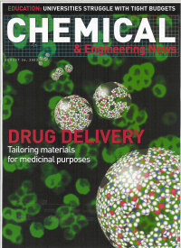 The project with the MNiMBS on dendrimer-based drug transport agents was highlighted on the cover of C&EN News (August 26, 2002).